
Cystoid Macular Edema (CME): Causes, Symptoms & Treatment
If your central vision has turned blurry or straight lines suddenly look wavy — especially in the weeks after a cataract surgery — fluid buildup in the macula could be the reason. Cystoid macular edema sounds alarming, but it's one of the most treatable retinal conditions when caught early. Here's exactly what it is, why it happens, and what treatment actually looks like.
Quick Answer
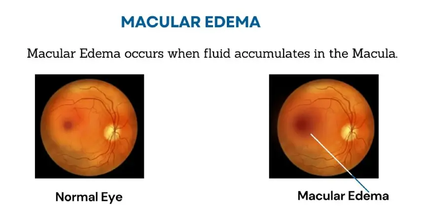
Cystoid macular edema (CME) occurs when fluid leaks from tiny blood vessels and pools in cyst-like pockets within the macula — the part of the retina responsible for sharp, central vision. This causes blurred or distorted central vision, faded colours, and difficulty with detailed tasks like reading or recognising faces. The single most common trigger is cataract surgery, where it's specifically called Irvine-Gass syndrome; other causes include diabetes, uveitis, retinal vein occlusion, and certain medications. Most cases, especially those following cataract surgery, improve significantly or resolve completely with anti-inflammatory drops, injections, or time.
Key Takeaways
- CME happens when a breakdown in the eye's blood-retinal barrier lets fluid pool in cyst-like spaces within the macula.
- Cataract surgery is the single most common trigger, known specifically as Irvine-Gass syndrome, typically appearing 4-10 weeks after surgery.
- Other causes include diabetes, uveitis, retinal vein occlusion, epiretinal membrane, and certain eye medications.
- The classic symptom is blurred or wavy central vision — peripheral vision is usually unaffected, and some people have no symptoms at all.
- OCT scanning and fluorescein angiography are the key diagnostic tools, often revealing a distinctive "honeycomb" or "petaloid" pattern.
- Post-surgical CME resolves on its own in most cases within about six months; other causes may need more active, ongoing treatment.
Figures reflect general clinical research and can vary based on individual risk factors and surgical complexity.
What Exactly Is Cystoid Macular Edema?
Cystoid macular edema occurs when fluid leaks out of tiny blood vessels in and around the macula — the small, central part of the retina responsible for the sharp, detailed vision you use for reading, driving, and recognising faces. Normally, a protective barrier (the blood-retinal barrier) keeps fluid from the surrounding blood vessels contained. When this barrier is disrupted — by surgery, inflammation, or disease — fluid pools into small, cyst-like pockets within the retina's layers, causing the macula to swell and thicken. This swelling distorts the delicate structure of the macula, which is exactly what blurs and distorts central vision.
Why Is CME So Closely Linked to Cataract Surgery?
When cystoid macular edema develops after cataract surgery, it has a specific name: Irvine-Gass syndrome (also called pseudophakic CME). It's actually one of the most common causes of reduced vision following an otherwise successful cataract surgery, thought to result from post-surgical inflammation temporarily disrupting the blood-retinal barrier. It typically appears around 4 to 10 weeks after surgery. With modern small-incision techniques like phacoemulsification, the risk of clinically noticeable CME (the kind that actually affects vision) is low — roughly 1-2% in routine, uncomplicated cases. The reassuring part: the large majority of these cases resolve on their own, often within about six months, sometimes helped along with a short course of anti-inflammatory drops.
What Causes Cystoid Macular Edema?
Cataract or Other Eye Surgery
Most CommonPost-surgical inflammation can temporarily disrupt the blood-retinal barrier, leading to Irvine-Gass syndrome, as explained above. This is the single most frequent cause of CME overall.
Diabetes and Diabetic Retinopathy
CommonChronic high blood sugar damages the walls of retinal blood vessels over time, causing them to leak fluid into the macula. See our guide to diabetic retinopathy.
Retinal Vein Occlusion
CommonA blocked retinal vein raises pressure in the surrounding blood vessels, forcing fluid to leak out into nearby retinal tissue, including the macula.
Uveitis (Eye Inflammation)
CommonOngoing inflammation inside the eye is a well-recognised trigger for CME, since it directly disrupts the same blood-retinal barrier that normally keeps fluid contained. Learn more about our uveitis clinic.
Epiretinal Membrane
Less CommonA thin layer of scar tissue on the macula's surface can pull on and distort the retina, sometimes triggering fluid accumulation underneath.
Retinitis Pigmentosa
Less CommonThis inherited retinal condition is sometimes associated with CME as one of its recognised complications, even independent of surgery or diabetes.
Certain Medications
Less CommonProstaglandin analog eye drops (commonly used for glaucoma) and niacin (vitamin B3) supplements have both been linked to CME in some patients, usually reversible on stopping the medication under medical guidance.
What Are the Symptoms of CME?
The bending of straight lines is a specific symptom doctors call metamorphopsia, and it's one of the more distinctive clues pointing toward macular involvement. It's worth knowing that peripheral (side) vision is usually completely unaffected by CME, and some people with mild, early CME have no noticeable symptoms at all — which is one reason it's sometimes first picked up on a routine post-surgical scan rather than because the patient reported a problem.
When Should You Seek Immediate Care?
Most CME develops gradually and isn't an emergency, but treat these as urgent:
- Sudden loss of vision, rather than a gradual change
- Severe eye pain
- Persistent new flashes of light or floaters
- Any significant, rapid change in vision, especially after recent eye surgery
How Is Cystoid Macular Edema Diagnosed?
Dilated Eye Exam
Your eye specialist uses dilating drops to widen the pupil and examines the retina directly for signs of swelling or fluid accumulation.
Optical Coherence Tomography (OCT)
A painless, non-invasive scan that produces detailed cross-section images of the retina, often revealing the classic "honeycomb" pattern of fluid-filled cystic spaces.
Fluorescein Angiography (FA)
A fluorescent dye is injected into a vein in your arm, and photos of the retina are taken as it circulates through, revealing blood vessel leakage in a distinctive "petaloid" (flower-petal) pattern — considered the gold standard for confirming CME.
What Are the Treatment Options for Cystoid Macular Edema?
Treatment depends on the underlying cause and how severe the swelling is. Common options include:
Topical Anti-Inflammatory Drops
NSAID or steroid eye drops are often the first step, particularly for CME following cataract surgery, helping to calm inflammation and reduce fluid leakage.
Intravitreal Injections
Anti-VEGF or corticosteroid medication injected directly into the eye targets the site of fluid buildup, reducing swelling and improving vision. The injection itself is quick and performed under numbing drops so it isn't painful.
Laser Photocoagulation
A focused laser can seal specific leaking blood vessels in the retina, used selectively depending on the pattern and cause of leakage.
Vitrectomy
In select, more complex cases, this surgical procedure removes the vitreous gel (replacing it with a saline solution), which can relieve traction contributing to the swelling.
Managing the Underlying Condition
When CME is linked to diabetes, uveitis, or high blood pressure, controlling that condition effectively is central to preventing further swelling and protecting vision long-term.
How Is Age-Related Macular Degeneration Different From Macular Edema?
| Aspect | Age-Related Macular Degeneration (AMD) | Cystoid Macular Edema |
|---|---|---|
| Definition | Progressive degeneration of the macula | Fluid accumulation in the macula |
| Typically affects | Adults aged 50 and above | Any age, depending on cause |
| Types | Dry AMD (slow) and Wet AMD (rapid) | Varies by underlying cause |
| Common causes | Ageing, genetics, smoking, diet | Cataract surgery, diabetes, vein occlusion, uveitis |
| Treatment | Risk-factor management, injections, laser | Treat underlying cause, drops, injections |
What Is the Long-Term Outlook for CME?
The outlook for cystoid macular edema depends heavily on its cause. CME that follows cataract surgery has a genuinely encouraging outlook — the vast majority of cases resolve spontaneously or with a short course of drops within about six months, with little to no lasting effect on vision. CME linked to an ongoing condition like diabetes, uveitis, or retinal vein occlusion tends to need more active, sometimes long-term management, and can recur if the underlying condition isn't well controlled. Across all causes, the same pattern holds true: the earlier CME is diagnosed and treated, the better the visual outcome tends to be — which is exactly why routine follow-up after cataract surgery, and regular monitoring for at-risk patients, matters so much.
Can Cystoid Macular Edema Be Prevented?
- Attend all recommended post-surgical follow-ups — post-cataract CME is often caught on a routine check before you'd notice any symptoms yourself
- Keep chronic conditions well controlled — working closely with your physician to manage diabetes or high blood pressure meaningfully lowers your risk
- Choose a healthy lifestyle — a balanced diet rich in fruits and vegetables, regular physical activity, and not smoking all support overall eye health
- Protect your eyes — UV-blocking sunglasses and appropriate protective eyewear during sports help prevent injury-related complications
- Get regular eye exams — routine check-ups help catch early, often symptom-free changes before they affect your vision
Dr. Rajeev Jain, Director, Save Sight Centre
"Patients are often surprised to hear that mild macular swelling after cataract surgery is fairly common and usually resolves on its own — the word 'edema' sounds far more alarming than the reality for most cases. What actually matters is routine follow-up, so if it is one of the cases that needs treatment, we catch it early and get ahead of it, rather than letting it linger."
Frequently Asked Questions About Cystoid Macular Edema
The best treatment depends on the individual case and its underlying cause. It could include topical anti-inflammatory drops, intravitreal injections, laser therapy, or treating an underlying condition like diabetes. Your retina specialist chooses the approach based on your specific diagnosis and how severe the swelling is.
The duration varies widely. CME following cataract surgery often resolves on its own within a few months in the majority of cases, sometimes helped along with anti-inflammatory drops. CME linked to diabetes, uveitis, or vein occlusion may take longer and need ongoing treatment. Regular follow-ups with your eye doctor help track progress and adjust the plan as needed.
There isn't one universal drug — the choice depends on the cause. Topical NSAID and steroid eye drops are often tried first, especially after cataract surgery. For more persistent or vision-threatening cases, intravitreal anti-VEGF or corticosteroid injections directly into the eye are commonly used to reduce swelling and improve vision.
For mild, post-surgical CME, topical anti-inflammatory drops are usually tried first. For more significant or vision-affecting swelling, intravitreal injections are often the first-line treatment, delivering medication directly to where the fluid is accumulating.
It may not always be permanently cured, especially when linked to an ongoing condition like diabetes, but it can very often be effectively managed and significantly improved. Early detection and the right treatment — injections, drops, or laser therapy — lead to much better outcomes, and regular eye check-ups help keep the treatment plan on track.
Irvine-Gass syndrome is simply the name given to cystoid macular edema that develops after cataract surgery — it's the same condition, just linked to a specific cause. It's actually one of the most common causes of CME overall, typically appearing around 4 to 10 weeks after surgery, and resolving on its own in the vast majority of cases within about six months.
With modern small-incision cataract surgery techniques like phacoemulsification, clinically noticeable CME (the kind that actually affects vision) occurs in roughly 1-2% of routine cases. If doctors look specifically with OCT scanning, mild, often symptom-free swelling can be picked up somewhat more often, but most of these cases resolve without any lasting effect on vision.
No. Most cases, particularly those following cataract surgery, improve substantially or resolve completely with time and appropriate treatment. Vision loss becomes more of a risk with chronic, long-standing, or repeatedly recurring CME that goes untreated, which is why timely diagnosis and follow-up matter so much.
Yes, it can recur, particularly when the underlying cause is an ongoing condition like diabetes, uveitis, or a retinal vein occlusion. This is why patients with these conditions are usually followed up regularly even after their CME has resolved, so any recurrence can be caught and treated early.
They overlap but aren't identical. Diabetic macular edema is macular swelling specifically caused by diabetes, and it can show the same cystoid (cyst-like) pattern seen in CME from other causes. In other words, diabetic macular edema is one possible cause of a cystoid pattern, while "cystoid macular edema" describes the pattern of swelling itself, regardless of what's causing it.
Noticing Blurry or Wavy Central Vision?
Get an accurate diagnosis and expert retina care at Save Sight Centre, Adarsh Nagar, North Delhi. NABH & ISO accredited · 15+ years · 1,70,000+ patients treated.
Disclaimer: The information on this site should not be considered medical advice. Please consult a doctor for determining the best procedure as well as the most appropriate treatment for your eyes. Results may vary from person to person and we do not guarantee similar results for everyone.