
9 Corneal and External Eye Diseases: Symptoms, Causes & Treatment
The cornea is the tiny, transparent window your entire vision depends on — so when something's wrong with it, or with the surface around it, it's worth knowing exactly what you're dealing with. Here's a plain-language guide to the most common corneal and external eye conditions: what they feel like, why they happen, and how they're actually treated.
Quick Answer
Corneal and external eye diseases are conditions affecting the cornea (the eye's clear front surface) or the structures around it — the conjunctiva, eyelids, tear film, and sclera. Common corneal conditions include keratitis, herpes simplex keratitis, keratoconus, and Fuchs' dystrophy. Common external conditions include dry eye disease, pterygium, allergic conjunctivitis, and rarer but serious conditions like Stevens-Johnson syndrome and ocular cicatricial pemphigoid. Most respond very well to treatment — from eye drops and specialty contact lenses to corneal cross-linking or a corneal transplant — especially when caught early.
Key Takeaways
- The cornea provides 65-75% of your eye's focusing power and has no blood vessels, which is what keeps it transparent.
- Corneal diseases affect the cornea directly; external eye diseases affect the conjunctiva, eyelids, tear film, or sclera around it.
- Keratitis, keratoconus, dry eye disease, and pterygium are among the most common conditions seen in everyday practice.
- Fuchs' dystrophy and keratoconus are progressive but manageable when caught early — a routine eye exam often catches them before symptoms worsen.
- Left untreated, several of these conditions can cause permanent vision loss, but most respond very well to timely treatment.
- Good contact lens hygiene, UV protection, and regular eye exams meaningfully lower your risk across almost all of these conditions.
Figures are general clinical guidance and can vary from person to person; a proper eye exam is needed for individual diagnosis.
What Is the Cornea and Why Does It Matter?
The cornea is the clear, dome-shaped outer layer covering your iris (the coloured part of your eye) and pupil. Unlike most tissues in your body, it has no blood vessels — which is exactly what keeps it transparent and able to bend (refract) light properly for clear vision. It does three essential jobs: it provides roughly 65-75% of your eye's total focusing power, it acts as a protective barrier against germs, dust and injury, and it helps maintain your eye's shape against internal pressure.
What Are the Layers of the Cornea?
The cornea isn't a single sheet of tissue — it's made of five distinct layers, each with its own job:
The outermost epithelium keeps out dust, debris and bacteria while absorbing nutrients from your tears. Beneath it, the tough Bowman's layer protects the deeper tissue. The stroma makes up around 90% of the cornea's thickness and gives it strength and clarity. The thin but sturdy Descemet's membrane shields against infection, and the innermost endothelium constantly pumps fluid out to keep the cornea from swelling — this is the layer that fails in Fuchs' dystrophy.
What's the Difference Between Corneal and External Eye Diseases?
Corneal diseases affect the cornea itself — the clear window at the front of your eye. External eye diseases affect the structures surrounding it: the conjunctiva (the membrane covering the white of your eye), the eyelids, the tear film, and the sclera. The two categories often overlap in practice, since problems on the eye's surface can spread to or affect the cornea if left untreated — which is exactly why both types need proper attention.
What Are the Most Common Corneal Diseases?
These conditions affect the cornea directly:
Keratitis (Corneal Ulcer)
InfectiousSymptoms: Red, painful eyes, blurry or hazy vision, watery or sticky discharge, light sensitivity.
Causes: Bacterial, viral or fungal infection, dirty or old contact lenses, eye injury, or swimming without goggles.
Treatment: Antibiotic, antifungal or antiviral drops as prescribed, temporarily stopping contact lens wear, and regular check-ups. Never self-medicate — contact your eye specialist right away.
Herpes Simplex Keratitis
Viral, RecurringSymptoms: Eye redness, mild pain or irritation, watery discharge, blurred vision that can worsen with flare-ups.
Causes: The herpes simplex virus (HSV-1) — the same virus behind cold sores. Stress, low immunity or illness can trigger a flare-up.
Treatment: Antiviral eye drops or oral medication, lubrication for comfort, and regular monitoring to prevent recurrence and complications.
Keratoconus
ProgressiveSymptoms: Blurry or wavy vision, light sensitivity, frequent changes in glasses power, poor night vision, discomfort with soft contact lenses.
Causes: Genetics, chronic eye rubbing, and allergic conditions. It usually starts in the teenage years or early adulthood.
Treatment: Custom glasses or contact lenses, corneal collagen cross-linking (CXL) to halt progression, INTACS ring segments, or a corneal transplant in advanced cases.
Fuchs' Endothelial Dystrophy
ProgressiveSymptoms: Blurred vision that's often noticeably worse in the morning, glare, gradual worsening over years, occasional pain in advanced stages.
Causes: Progressive loss of the endothelial cells that pump fluid out of the cornea, causing it to swell. Usually appears after age 50 and often runs in families.
Treatment: Lubricating drops or ointments for mild cases; a partial-thickness corneal transplant (DSEK or DMEK) for advanced disease.
What Are the Most Common External Eye Diseases?
These conditions affect the conjunctiva, eyelids, tear film, or sclera around the cornea:
Dry Eye Disease
Very CommonSymptoms: Burning or stinging, a gritty feeling, red or tired eyes, blurred vision that worsens while reading or on screens.
Causes: Long screen time without blinking, ageing, certain medications, or dry, polluted environments.
Treatment: Artificial tears and gels, anti-inflammatory drops, punctal plugs, and better blinking habits during screen use.
Pterygium
Benign GrowthSymptoms: A visible pink, triangular growth on the eye, irritation, redness, blurred vision if it extends onto the cornea.
Causes: UV sun exposure, wind, dust, and dry climates causing chronic irritation.
Treatment: Lubricating drops, anti-inflammatory medication, or surgical removal with a graft to prevent regrowth if it affects vision.
Allergic Conjunctivitis
SeasonalSymptoms: Itchy, red eyes, swollen eyelids, watering or discharge, a burning sensation.
Causes: Pollen, dust, pet hair, pollution, smoke, or contact lens sensitivity.
Treatment: Anti-allergy eye drops, lubricants to flush out allergens, cold compresses, and avoiding known triggers.
Stevens-Johnson Syndrome
Rare, SevereSymptoms: Eye redness and pain, blisters or sores on the eyelids, dry eyes, vision loss in serious cases.
Causes: A severe allergic reaction to medication, or sometimes viral or bacterial infection.
Treatment: Specialised lubricating and healing drops, amniotic membrane therapy, surgical repair if needed, and coordinated care with dermatologists.
Ocular Cicatricial Pemphigoid
Rare, AutoimmuneSymptoms: Red, irritated eyes, dryness and discomfort, progressive scarring of the eyelids or conjunctiva, gradual vision loss.
Causes: An autoimmune response where the body attacks its own eye surface tissue.
Treatment: Immunosuppressive medication, lubricating and anti-inflammatory drops, and eyelid correction surgery where needed, with ongoing monitoring.
Which Corneal Disease Matches Your Symptoms?
| If you notice... | It could suggest |
|---|---|
| Sudden pain, redness, discharge | Keratitis or corneal ulcer |
| Recurring one-eye irritation, past cold sores | Herpes simplex keratitis |
| Frequent glasses power changes, distorted vision | Keratoconus |
| Morning blur that improves through the day | Fuchs' dystrophy |
| Burning, grittiness, screen-related blur | Dry eye disease |
| Visible pink growth on the eye surface | Pterygium |
| Itching, swelling, seasonal pattern | Allergic conjunctivitis |
This is a general guide, not a diagnosis. Several conditions share overlapping symptoms — a proper eye exam is the only way to know for certain.
When Should You See an Eye Specialist Immediately?
Your eyes are precious, and early detection genuinely saves vision. See a specialist right away if you notice:
- Ongoing redness or eye pain that isn't settling
- Sudden changes in vision or new blurry spots
- Increased sensitivity to light
- An eye injury, or something stuck in your eye
- Frequent eye infections or a corneal ulcer that isn't healing
What Is a Corneal Transplant and When Is It Needed?
A corneal transplant (keratoplasty) replaces damaged corneal tissue with healthy donor tissue, and is considered when scarring, thinning, or swelling has progressed too far for other treatments to restore useful vision. Not all transplants are the same — the technique depends on which layer of the cornea is affected:
| Type | What It Replaces | Typically Used For |
|---|---|---|
| Penetrating Keratoplasty (PK) | Full corneal thickness | Severe scarring across all layers |
| DALK | Front and middle layers only | Advanced keratoconus with a healthy endothelium |
| DSEK / DMEK | Only the innermost endothelium | Fuchs' dystrophy and endothelial failure |
Donor corneas don't need to match your blood type, and modern partial-thickness techniques (like DSEK and DMEK) have significantly shortened recovery time compared to older full-thickness surgery.
How Are Corneal Diseases Diagnosed?
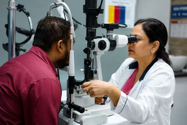
Slit-Lamp Examination
A magnified, detailed view of the cornea and surrounding tissue — the starting point for almost every diagnosis.
Corneal Topography
Maps the curvature of your cornea in detail; essential for diagnosing and tracking keratoconus.
Tear Film Break-Up Time (TBUT)
Measures how stable your tear film is, a key test in diagnosing dry eye disease.
OCT (Optical Coherence Tomography)
Produces high-resolution cross-section images of the corneal layers and nearby tissue.
Microbiological Testing
Identifies the exact bacteria, virus, fungus, or parasite responsible for an infection, guiding precise treatment.
How Can You Prevent Corneal and External Eye Problems?
- Clean and handle contact lenses properly — never sleep in them unless your doctor advises it
- Wear UV-protective sunglasses outdoors
- Follow the 20-20-20 rule to reduce screen-related strain
- Avoid rubbing your eyes, especially with unwashed hands
- Drink plenty of water to keep your eyes naturally lubricated
- Use lubricating eye drops in dry or windy environments
- Eat a diet rich in omega-3 fatty acids and antioxidants to support eye health
Tip: If you notice any of the warning symptoms above, don't wait it out — contact an eye specialist without delay.
Dr. Rajeev Jain, Director, Save Sight Centre
"The cornea is unforgiving of neglect but very forgiving of early treatment. We see patients who ignored a gritty, red eye for weeks thinking it was 'just strain,' and patients who came in the same day and walked out with a simple course of drops. The difference in outcome is almost always about timing, not severity — which is exactly why I tell every patient: when in doubt, get it checked."
Frequently Asked Questions About Corneal & External Eye Diseases
Keratitis (corneal ulcer), herpes simplex keratitis, keratoconus, and Fuchs' endothelial dystrophy are among the most common. They range from infections that develop quickly and need urgent treatment, to slow, progressive conditions like keratoconus and Fuchs' dystrophy that are usually caught during a routine eye exam.
External eye diseases affect the structures around the cornea rather than the cornea itself — the conjunctiva, eyelids, tear film, and sclera. Common examples include dry eye disease, pterygium, allergic conjunctivitis, and rarer but serious conditions like Stevens-Johnson syndrome and ocular cicatricial pemphigoid.
Redness, pain, blurry or distorted vision, watering, light sensitivity, a gritty or foreign-body sensation, and any visible growth or discharge are all worth paying attention to. Symptoms that are sudden, severe, or don't improve within a day or two should be checked by an eye specialist promptly.
Yes, if left untreated. Severe keratitis, advanced keratoconus, Fuchs' dystrophy, and autoimmune conditions like ocular cicatricial pemphigoid can all lead to significant, sometimes permanent, vision loss. The encouraging part is that most of these conditions respond very well to treatment when caught early.
Diagnosis typically starts with a slit-lamp examination for a magnified view of the cornea, along with vision testing. Depending on the suspected condition, doctors may add corneal topography to map its curvature, tear film break-up time for dry eye, OCT imaging for detailed cross-sections, or microbiological testing if an infection is suspected.
Treatment depends entirely on the condition — options range from medicated eye drops and lubricating tears, to procedures like corneal collagen cross-linking for keratoconus, to a corneal transplant for advanced disease or scarring. Your eye specialist will match the treatment to your specific diagnosis and how far it has progressed.
Practice good contact lens hygiene, never sleep in your lenses unless advised, wear UV-protective sunglasses outdoors, follow the 20-20-20 rule to reduce screen strain, avoid rubbing your eyes, stay hydrated, and get regular eye check-ups so problems are caught before they progress.
See an eye specialist if you notice ongoing redness or pain, sudden vision changes or blurry spots, increased light sensitivity, an eye injury or foreign object, or infections that keep recurring or won't heal. Early evaluation is the single biggest factor in protecting your long-term vision.
Fuchs' dystrophy is a slow, progressive condition where the innermost layer of the cornea loses its ability to pump out excess fluid, causing the cornea to swell and vision to blur, often noticeably worse in the morning. Mild cases are managed with lubricating drops or ointments; advanced cases are treated with a partial corneal transplant (DSEK or DMEK).
Keratoconus can't be reversed, but its progression can be halted, most effectively with corneal collagen cross-linking, especially when started early. Vision can then be rehabilitated with glasses, specialty contact lenses, or in advanced cases, a corneal transplant, which restores functional vision for the vast majority of patients.
The procedure itself is done under anaesthesia, so it isn't painful, though some discomfort during recovery is normal and manageable with medication. Corneal transplants have a high success rate — donor corneas don't need to match blood type, and modern partial-thickness techniques have significantly improved recovery time and outcomes compared to older full-thickness surgery.
No, a pterygium is a benign, non-cancerous growth. It's usually harmless and only needs treatment if it causes persistent irritation, redness, or starts to grow onto the cornea enough to affect vision, in which case it can be surgically removed.
Not Sure What's Going On With Your Eyes?
Get an accurate diagnosis with advanced corneal diagnostics at Save Sight Centre, Adarsh Nagar, North Delhi. NABH & ISO accredited · 15+ years · 1,70,000+ patients treated.
Disclaimer: For general awareness only, not medical advice. Please consult an eye specialist at Save Sight Centre for a proper diagnosis and treatment plan tailored to your eyes. Results vary by person. Figures cited are approximate and based on general clinical patterns.