

 +91-7873-222-000
 WhatsApp

 Book Appointment

Retinal Hemorrhage: Causes, Symptoms & Treatment
A retinal hemorrhage happens when the delicate blood vessels feeding your retina break or leak — and because the retina is what actually converts light into the images you see, even small amounts of bleeding can blur or distort your vision. The encouraging news: caught early, the vast majority of cases respond very well to treatment. Here's what causes it, how to recognise it, and what treatment actually looks like.

Quick Answer
A retinal hemorrhage occurs when blood vessels within the retina — the light-sensitive tissue at the back of the eye — break or leak blood, which can blur vision, cause floaters, or in severe cases, lead to sudden vision loss. The most common causes are diabetes and high blood pressure, which gradually weaken retinal blood vessels; other causes include trauma, retinal vein occlusion, clotting disorders, and age-related macular degeneration. Small hemorrhages often clear on their own within a few weeks; larger ones typically need laser treatment, anti-VEGF injections, or in select cases, surgery to protect vision.
Key Takeaways
- Diabetes and high blood pressure are the two leading causes of retinal hemorrhage worldwide.
- Hemorrhages are classified by location and depth — flame-shaped, dot-and-blot, preretinal, and subretinal — each pointing toward different likely causes.
- A retinal hemorrhage is different from a vitreous hemorrhage (bleeding into the gel in front of the retina), though the two are closely related and can occur together.
- Sudden floaters or a shadow across your vision needs prompt evaluation, since a hemorrhage can sometimes hide a retinal tear or detachment underneath.
- Small hemorrhages often resolve on their own; larger ones respond well to laser therapy, anti-VEGF injections, or vitrectomy surgery.
- Managing diabetes and blood pressure is the single most effective way to reduce your long-term risk.
Figures reflect general clinical research and can vary widely based on the underlying cause and severity.
What Is a Retinal Hemorrhage?
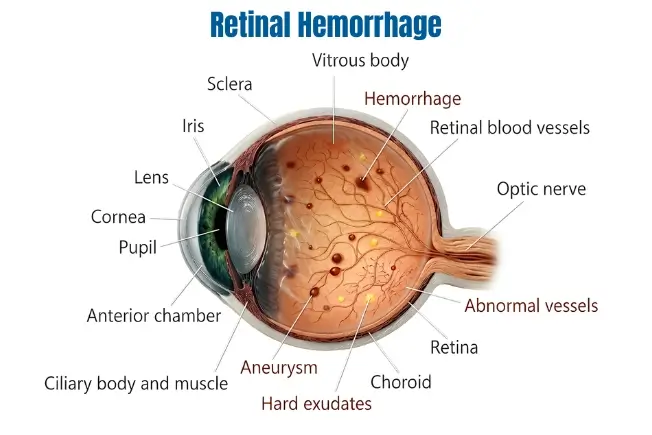
A retinal hemorrhage occurs when the blood vessels within the retina — the light-sensitive layer lining the back of your eye — break or leak blood. Since the retina's job is to convert light into the signals your brain interprets as images, blood pooling within or in front of it directly interferes with this process, blurring or distorting vision depending on the size and location of the bleed. It's most common in people with diabetes or high blood pressure, though trauma, blood-clotting disorders, and several other conditions can cause it too. The reassuring part: with early detection and the right treatment, the vast majority of cases preserve useful vision.
Is a Retinal Hemorrhage the Same as a Vitreous Hemorrhage?
They're closely related but not identical, and the distinction matters. A retinal hemorrhage is bleeding within the layers of the retina itself. A vitreous hemorrhage is bleeding into the vitreous — the clear, gel-like substance that fills the space between your lens and your retina. The two often occur together, since blood from a retinal bleed can break through into the vitreous, and both share similar underlying causes like diabetic retinopathy and trauma. Vitreous hemorrhage tends to cause more sudden, dramatic vision loss, since it clouds the entire path light must travel through the eye rather than affecting just the retina's surface — it has an estimated annual incidence of around 7 cases per 100,000 people, making it one of the more common causes of sudden, painless vision loss seen in eye emergency clinics.
What Are the Types of Retinal Hemorrhage?
Doctors classify retinal hemorrhages by their shape, position, and depth within the retinal layers — this classification helps predict the likely cause and guide treatment.
Flame-Shaped Hemorrhages
SuperficialAppear as streak-like marks in the nerve fibre layer, commonly linked to high blood pressure. These typically resolve more quickly than deeper hemorrhages.
Dot-and-Blot Hemorrhages
Deeper LayerRound, compact spots found in the deeper, inner retinal layers, most often linked to diabetes. Patients with this pattern usually need long-term blood sugar management to prevent recurrence.
Layered (Multi-Level) Hemorrhages
More SeriousAffect multiple retinal levels simultaneously and often signal a more significant underlying vascular problem, such as retinal vein occlusion.
Preretinal Hemorrhages
In Front of RetinaDevelop between the retina and the vitreous gel. Because of their position directly along the visual pathway, they tend to obstruct vision more severely than deeper hemorrhages.
Subretinal Hemorrhages
Beneath RetinaOccur beneath the retina itself and primarily distort central vision, since they frequently involve the macula, the retina's central, detail-focused region.
Identifying the exact type helps your ophthalmologist predict recovery time more accurately — smaller hemorrhages often clear within weeks, while larger preretinal or subretinal hemorrhages sometimes need surgical treatment.
What Are the Symptoms of Retinal Hemorrhage?
Retinal hemorrhage isn't usually painful, though headaches can accompany the visual changes. It affects one eye more often than both, and symptoms can develop gradually or appear suddenly after an injury.
Floaters — dark spots, webs, or strings drifting across your field of vision — are one of the most common early signs. As bleeding progresses, some people notice a shadow slowly filling part of their visual field, similar to a curtain being drawn, or blind spots limiting their central focus (a pattern called a scotoma). In more severe cases, vision in the affected eye can be lost quite quickly. Diabetic patients sometimes notice symptoms flare specifically when blood sugar is poorly controlled. If these signs are ignored, scar tissue can eventually develop, causing more permanent vision changes.
What Causes Retinal Hemorrhage?
Diabetes
Leading CauseChronically high blood sugar weakens the fragile blood vessels in the retina over time, eventually causing them to leak or burst. See our guide to diabetic retinopathy.
High Blood Pressure (Hypertension)
Sustained high pressure puts excess strain on already fragile retinal blood vessels, eventually causing them to rupture.
Physical Trauma
Falls, sports injuries, or accidents involving direct impact to the eye or head can cause immediate retinal bleeding.
Retinal Vein Occlusion
A blockage in normal retinal blood flow causes pressure to build up behind the obstruction, eventually rupturing the vessel wall.
Blood-Clotting Disorders
Systemic conditions that interfere with normal clotting, or blood-thinning medications, can make retinal vessels more prone to bleeding.
Valsalva Retinopathy (Straining)
Often OverlookedA sudden, forceful strain — heavy lifting, intense vomiting, forceful coughing, or difficult childbirth — sharply raises pressure in the chest and abdomen, which can transmit back to the eye and rupture a retinal vessel, even in someone with otherwise healthy eyes.
Age-Related Macular Degeneration
Vascular changes linked to AMD make the retina more vulnerable to bleeding, particularly after age 60.
Autoimmune Diseases and Rare Conditions
Some autoimmune conditions directly inflame retinal blood vessels. Rare systemic conditions such as leukaemia or infectious endocarditis can also present with scattered retinal hemorrhages.
Why Might a Hemorrhage Be Hiding a More Serious Problem?
This is one of the most important things to understand about sudden retinal or vitreous bleeding: the blood itself can obscure your doctor's view of the retina, potentially hiding a retinal tear or detachment underneath — arguably the most concerning cause of a spontaneous hemorrhage, since a missed tear or detachment carries a real risk of permanent vision loss if not treated promptly. This is exactly why any sudden onset of floaters, a shadow, or vision loss needs a proper dilated eye exam as soon as possible, even if the hemorrhage itself later turns out to be minor — your ophthalmologist needs to actively rule out what might be hiding behind it, sometimes using ultrasound imaging if the blood is too dense to see through directly.
How Is Retinal Hemorrhage Diagnosed?
Dilated Fundus Examination
Widening the pupil allows your ophthalmologist a full, detailed view of the retina to assess the location, size, and pattern of bleeding.
Optical Coherence Tomography (OCT)
Produces detailed cross-section images of the retina, helping identify exactly which layers are affected.
Fluorescein Angiography
A dye is injected into the bloodstream to highlight leaking or abnormal blood vessels feeding the retina.
B-Scan Ultrasound
Used when a dense hemorrhage blocks a clear direct view of the retina, helping confirm whether a retinal tear or detachment is present underneath.
What Are the Treatment Options for Retinal Hemorrhage?
Non-surgical options are tried first in most cases:
Observation
Many small retinal hemorrhages resolve spontaneously within a few weeks; your retina specialist will schedule regular follow-ups to confirm this is happening safely.
Laser Photocoagulation
Precisely seals leaking blood vessels to prevent further bleeding, usually a brief, well-tolerated in-office procedure.
Anti-VEGF Injections
Administered directly into the eye, typically every four to six weeks initially, to suppress abnormal vessel growth and reduce leakage.
Surgical options are reserved for more severe or non-resolving cases:
Vitrectomy
Removes accumulated blood, vitreous gel, and scar tissue that may be causing traction on the retina — often needed for dense, non-clearing hemorrhages.
Managing the Underlying Cause
Oral medications to control diabetes, antihypertensive treatment to stabilise blood pressure, or anticoagulation adjustments for clotting disorders — all protect the remaining healthy blood vessels from further damage.
What Is the Outlook for Retinal Hemorrhage?
Outcomes depend heavily on the size, location, and underlying cause of the hemorrhage, and on how quickly treatment begins. Most patients on anti-VEGF injections need roughly four to six injections a year, adjusted based on response. Laser treatments are brief and cause minimal discomfort. Vitrectomy surgery typically allows recovery over a few months, supported by topical medication during healing. Across all approaches, combining treatments — managing the systemic cause alongside direct eye treatment — consistently gives the best long-term results.
How Can You Reduce Your Risk of Retinal Hemorrhage?
- Keep blood sugar well controlled — consistent management through diet, medication, and regular monitoring meaningfully lowers your risk if you have diabetes
- Monitor and manage blood pressure — both at home and during regular medical check-ups
- Avoid smoking — smoking accelerates damage to blood vessels throughout the body, including the retina
- Be mindful of sudden straining — where possible, avoid holding your breath during heavy lifting, and get persistent vomiting or coughing checked and treated
- Wear protective eyewear — during sports or in occupations with a risk of eye injury
- Get an annual dilated eye exam — especially important if you have diabetes, hypertension, or a family history of retinal vein occlusion, since this catches silent changes before they progress
- Discuss blood-thinning medication with your doctor — if you're on anticoagulants, regular monitoring helps balance their necessary benefits against bleeding risk
What Specialist Care Is Available at Save Sight Centre?
Our retina specialists at Save Sight Centre, Delhi, manage retinal hemorrhage cases routinely, using the latest laser technology and intravitreal injection therapy to support timely healing. Comprehensive diagnostic imaging — including OCT, fluorescein angiography, and B-scan ultrasound where needed — helps identify underlying systemic causes efficiently. Our team works closely with diabetes and hypertension management specialists to ensure the root cause is addressed alongside direct eye treatment, and individualised treatment plans are built around each patient's specific hemorrhage type and overall health.
Dr. Rajeev JainDirector · Retina, Cataract, LASIK & Uvea Specialist
"The patients who do best are almost always the ones who came in the same week they noticed floaters or a shadow, rather than waiting to see if it would go away. A retinal hemorrhage that looks alarming can sometimes turn out to be quite manageable — and one that seems mild can occasionally be hiding something more serious behind it. Either way, a prompt exam is what tells us which situation we're actually dealing with."
Frequently Asked Questions About Retinal Hemorrhage
Diabetes and high blood pressure are responsible for the majority of retinal hemorrhages, since both gradually weaken the delicate blood vessels in the retina over time. High blood sugar and elevated vascular pressure often interact to produce the typical leakage patterns doctors see on examination.
Small retinal hemorrhages often improve without intervention within a few weeks as the body naturally reabsorbs the blood. Larger hemorrhages usually need treatment with laser therapy or injections to stop further bleeding and help the blood clear.
Cost varies considerably depending on the cause, severity, and treatment needed — simple monitoring costs far less than anti-VEGF injections, which in turn cost less than vitrectomy surgery. Your retina specialist can give you an accurate, personalised estimate once the type and extent of the hemorrhage has been assessed.
Extensive, untreated retinal hemorrhages can lead to permanent vision loss through scarring or retinal detachment. However, in the large majority of cases, laser therapy or injections given at an early stage are able to preserve useful vision, which is exactly why prompt evaluation matters so much.
Yes, it's very common in diabetic patients with long-standing or poorly controlled blood sugar. Annual retina check-ups are specifically designed to catch these early, often symptom-free changes before they progress to more severe vision loss.
A retinal hemorrhage is bleeding within the layers of the retina itself. A vitreous hemorrhage is bleeding into the vitreous — the clear, gel-like substance filling the space in front of the retina. The two are closely related and can occur together, since blood from a retinal bleed can break through into the vitreous, but a vitreous hemorrhage often causes more sudden and dramatic vision loss because it clouds the entire path light must travel through the eye.
Many smaller retinal hemorrhages heal completely with no lasting effect on vision, especially once the underlying cause (like blood pressure or blood sugar) is brought under control. Larger or recurrent hemorrhages can leave some residual scarring or vision changes, particularly if treatment is delayed, which is why early evaluation improves the odds of a full recovery.
Yes — this is a recognised cause called Valsalva retinopathy. A sudden, forceful strain, such as heavy lifting, intense vomiting, forceful coughing, or even difficult childbirth, sharply raises pressure in the chest and abdomen, which can transmit back to the delicate vessels in the eye and cause a hemorrhage, even in someone with otherwise healthy eyes.
Strict bed rest generally isn't required for most retinal hemorrhages, though your doctor may advise avoiding strenuous activity, heavy lifting, or anything that raises pressure in the head and chest while the hemorrhage is healing, particularly if straining was the original cause.
Treat sudden floaters, a shadow across your vision, or any abrupt vision change as urgent and get examined within 24 hours if possible. Even when a hemorrhage itself turns out to be minor, prompt examination is essential to rule out a retinal tear or detachment that could be hiding behind it, since these need to be treated quickly to protect your vision.
Noticing Sudden Floaters or Blurred Vision?
Get prompt retina evaluation and treatment at Save Sight Centre, Adarsh Nagar, North Delhi. NABH & ISO accredited · 15+ years · 1,70,000+ patients treated.
Disclaimer: The information on this site should not be considered medical advice. Please consult a doctor for determining the best procedure as well as the most appropriate treatment for your eyes. Results may vary from person to person and we do not guarantee similar results for everyone.